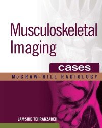
J. Tehranzadeh – Musculoskeletal Imaging Cases
3.450 ₽
Автор: J. Tehranzadeh
Название книги: Musculoskeletal Imaging Cases
Формат: PDF
Жанр: Медицина
Страницы: 909
Качество: Изначально компьютерное, E-book
“Over 70 international experts have submitted an excellent range of cases encompassing both essential concepts and encountered diagnoses. 3 Stars.”–Doody's Review Service
Musculoskeletal Imaging Cases offers an efficient and systematic approach to interpreting imaging of the musculoskeletal system. It delivers a one-of-a-kind collection of more than 370 cases with an emphasis on advanced imaging – including MRI, CT, US, and scintigraphy. The book's logical, easy-to-follow organization is designed for at-the-bench reference. The concise text, numerous multi-modality images, and helpful icons make looking up essential information fast and easy.
FEATURES:
More than 370 cases and 2000 multi-modality images
Each case highlights findings, differential diagnoses, and pearls, and contains multiple figures – some with colorful surgical specimens – to help you efficiently interpret and verify your diagnostic findings
Icons, a grading system depicting the spectrum of findings from common to rare, and the consistent chapter organization and structure provide ease-of-use for at-the-bench consultation
Special emphasis on the latest diagnostic modalities includes state-of-art depiction of image findings
Authorship from more than 70 contributing radiologists from the U.S. and around the globe provide a wealth of valuable cases
Описание
Musculoskeletal Imaging Cases от J. Tehranzadeh — это практическое пособие, которое помогает освоить интерпретацию изображений опорно-двигательного аппарата с помощью реальных клинических примеров. Книга построена в формате «случай — диагностика — разбор», что позволяет быстро связать рентгеновские снимки, КТ, МРТ и сцинтиграфию с конкретными заболеваниями и травмами.
Автор подробно разбирает патологии костей, суставов, мягких тканей, мышц и сухожилий. Особое внимание уделяется дифференциальной диагностике, типичным ошибкам и корреляции визуальных данных с клинической картиной. Издание охватывает как распространённые, так и редкие состояния в травматологии, ортопедии, ревматологии и онкологии.
- Рентгенологам и врачам лучевой диагностики, желающим улучшить навыки чтения снимков
- Травматологам-ортопедам и ревматологам для уточнения диагноза по визуализации
- Ординаторам и студентам старших курсов медицинских вузов, изучающим радиологию
- Всем специалистам, которые регулярно работают с МРТ и КТ опорно-двигательного аппарата
Только зарегистрированные клиенты, купившие данный товар, могут публиковать отзывы.
Похожие



Другие книги раздела «Лучевая диагностика и терапия (Радиология)»
- Alexander A. Bankier – Imaging in Transplantation
- Arjun Sahgal – Image-Guided Hypofractionated Stereotactic Radiosurgery
- Ingrid Reiser – Tomosynthesis Imaging
- Jeffrey S. Ross – Imaging in Spine Surgery (2017)
- Thomas Mang – CT Colonography
- Emanuele Neri – CT Colonography Atlas
- Vicente Sanchis-Alfonso – Atlas of the Patellofemoral Joint
- Thomas H. Berquist – Imaging of Orthopaedic Fixation Devices and Prostheses
- Sukru Mehmet Erturk – Teaching Atlas of Hepatobiliary and Pancreatic Imaging (2016)
- Sabine Weckbach – Incidental Radiological Findings
- Marc Lemmerling – Temporal Bone Imaging (2015)
- M. Oudkerk – Coronary Radiology (Second edition)
- Armanda Tatsas – Atlas of Radiologic Cytopathologic Correlations
- Ayman El-Baz – Multi Modality State-of-the-Art Medical Image Segmentation and Registration Methodologies (2 Volume set)
- Charles L. Epstein – Introduction to the Mathematics of Medical Imaging (Second edition)
- E. Coche – Comparative Interpretation of CT and Standard Radiography of the Chest
Все книги раздела «Лучевая диагностика и терапия (Радиология)» →
Смежные разделы: Медицина на испанском языке · Медицина на немецком языке · Медицина на французском языке · Анестезиология и Реаниматология · Акушерство и Гинекология · Ветеринария · Гастроэнтерология и Гепатология · Дерматология и косметология · Диетология и нутрициология · Иммунология и Аллергология · Кардиология · Лапароскопия и Эндоскопия
Рекомендуем также
- S. Paul – Clinical Cases in Skin Cancer Surgery and Treatment (2016)
- Y. Sakai – Laparoscopic Surgery for Colorectal Cancer (2016)
- V. Stearns – Novel Biomarkers in the Continuum of Breast Cancer (2016)
- A. Mathur – Nanotechnology in Cancer (2017)
- P. Rennert – Novel Immunotherapeutic Approaches to the Treatment of Cancer
- M. Baranska – Optical Spectroscopy and Computational Methods in Biology and Medicine (2014)
- A. Bondanza – Tumor Immunology (2016)
- H. Seligmann – DNA Replication. Current Advances (2016)
Отзывы
Отзывов пока нет.