
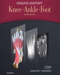
Julia R. Crim – Imaging Anatomy. Knee. Ankle. Foot (2017)
3.965 ₽
Автор: Julia R. Crim
Название книги: Imaging Anatomy. Knee. Ankle. Foot
Формат: PDF
Тема: Радиология, Анатомия
Страницы: 626
Качество: Изначально компьютерное, E-book
Designed to help you quickly learn or review normal anatomy and confirm variants, Imaging Anatomy: Knee, Ankle, Foot , by Dr. Julia R. Crim, provides detailed anatomic views of each major joint of the lower extremity. Ultrasound and 3T MR images in each standard plane of imaging (axial, coronal, and sagittal) accompany highly accurate and detailed medical illustrations, assisting you in making an accurate diagnosis. Comprehensive coverage of the knee, ankle, and foot, combined with an orderly, easy-to-follow structure, make this unique title unmatched in its field.
Includes all relevant imaging modalities, 3D reconstructions, and highly accurate and detailed medical graphics that illustrate the fine points of the imaging anatomy
Depicts common anatomic variants (both osseous and soft tissue) and covers imaging pitfalls as a part of its comprehensive coverage
Enables any structure in the lower extremity to easily be located, identified, and tracked in any plane for a faster, more accurate diagnosis
Provides richly labeled images with associated commentary as well as scout images to assist in localization
Explains uniquely difficult functional or anatomical regions of the lower extremity, such as posterolateral corner of knee, ankle ligaments, ankle tendons, and nerves of the lower extremity
Presents coronal and axial planes as both the right and left legs, on facing pages, making ultrasound/MR correlation even easier
Описание
Imaging Anatomy. Knee. Ankle. Foot — это подробный атлас по визуализационной анатомии нижней конечности от Julia R. Crim. Книга помогает врачам-радиологам, ортопедам и травматологам уверенно ориентироваться в сложных анатомических структурах коленного, голеностопного суставов и стопы при интерпретации МРТ, КТ и рентгеновских снимков.
Издание содержит высококачественные изображения с подробными анатомическими срезами в стандартных проекциях, акцентами на клинически значимых структурах, вариантах нормы, возрастных особенностях и типичных диагностических ловушках. Особое внимание уделено связкам, сухожилиям, хрящам, костным ориентирам и нейрососудистым пучкам.
- Радиологам, специализирующимся на опорно-двигательном аппарате
- Ортопедам-травматологам и хирургам стопы и голеностопа
- Рентгенологам, регулярно работающим с МРТ и КТ суставов
- Студентам старших курсов и ординаторам по специальностям «Травматология и ортопедия», «Лучевая диагностика»
Только зарегистрированные клиенты, купившие данный товар, могут публиковать отзывы.
Похожие




Другие книги раздела «Лучевая диагностика и терапия (Радиология)»
- Michael Riccabona – Pediatric Urogenital Radiology (2018)
- Sanjeev Agarwal – Radiology of Orthopedic Implants
- Ю. Лишманов – Национальное руководство по радионуклидной диагностике
- M. Runge – Radiologie De L’appareil Locomoteur (Французский язык)
- Б. Шах – Лучевая диагностика заболеваний молочной железы
- Paolo Russo – Handbook of X-ray Imaging
- Reinhard Schumacher – Fetal Radiology
- Axe Stabler – Handbuch Diagnostische Radiologie. Muskuloskelettales System (3 Тома, Немецкий язык)
- Nigel Raby – Notfallradiologie Unfallradiologie (Немецкий язык)
- In-One Kim – Radiology Illustrated. Pediatric Radiology
- Seung Hyup Kim – Uroradiology
- Xinwei Han – Airway Stenting in Interventional Radiology (2019)
- E. Scott Pretorius – Radiologia Secretos (Испанский язык)
- N. Reed Dunnick – Genitourinary Radiology (2018)
- Jeffrey Klein – Brant and Helms Fundamentals of Diagnostic Radiology (2019)
- Euclid Seeram – Digital Radiography (2019)
Все книги раздела «Лучевая диагностика и терапия (Радиология)» →
Смежные разделы: Медицина на испанском языке · Медицина на немецком языке · Медицина на французском языке · Анестезиология и Реаниматология · Акушерство и Гинекология · Ветеринария · Гастроэнтерология и Гепатология · Дерматология и косметология · Диетология и нутрициология · Иммунология и Аллергология · Кардиология · Лапароскопия и Эндоскопия
Рекомендуем также
- О. Швецова – Технология изготовления изделий по индивидуальным заказам
- R. Patel – Green Biocatalysis (2016)
- T. Jezierski – Canine Olfaction Science and Law (2016)
- M. Colecchia – Pathology of Testicular and Penile Neoplasms (2016)
- Н. Куликов – Водоснабжение (2016)
- C. Salgado – Aesthetic and Functional Surgery of the Genitalia
- A. Khan – Precision Molecular Pathology of Breast Cancer (2015)
- J. Rowley – Chromosomal Translocations and Genome Rearrangements in Cancer (2015)

Отзывы
Отзывов пока нет.