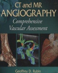
Geoffrey D. Rubin – CT and MR Angiography. Comprehensive Vascular Assessment
4.750 ₽
Автор: Geoffrey D. Rubin
Название книги: CT and MR Angiography. Comprehensive Vascular Assessment
Формат: PDF
Тема: Лучевая диагностика и терапия, МРТ и КТ
Страницы: 1339
Качество: Изначально компьютерное, E-book
Written by world-renowned experts in both CT angiography and MR angiography, this landmark work is the first comprehensive text on vascular imaging using CT and MR. It provides a balanced view of the capabilities of these modalities and practical guidelines for obtaining and interpreting images. More than 2,200 illustrations complement the text.
Chapters co-authored by CT and MR authorities cover imaging of all coronary and non-coronary arteries and veins. Each chapter details indications, imaging strategies, normal and variant anatomy, diseases, surgical management, and pitfalls. The authors compare the utility of CT and MR in specific clinical situations and discuss the role of conventional angiography and ultrasound where appropriate.
Описание
Книга CT and MR Angiography. Comprehensive Vascular Assessment Geoffrey D. Rubin — фундаментальное руководство по неинвазивной визуализации сосудов с помощью компьютерной и магнитно-резонансной томографии. Издание объединяет принципы получения изображений, основы сосудистых заболеваний и детальный разбор анатомии и патологии всех основных сосудистых регионов тела.
В первой части подробно разбираются физические принципы КТ- и МР-ангиографии, техника введения контрастных препаратов, постобработка данных и сравнение с традиционной ангиографией. Вторая часть посвящена атеросклерозу, тромбоэмболии, васкулитам и заболеваниям соединительной ткани. Третья часть — наиболее объёмная — содержит главы по анатомии и патологии церебральных, шейных, коронарных, лёгочных, аортальных, висцеральных, почечных, периферических артерий и вен, включая педиатрические аспекты.
- Рентгенологам и ангиологам, осваивающим КТ- и МР-ангиографию
- Кардиологам и сосудистым хирургам, нуждающимся в глубоком понимании возможностей современных методов визуализации
- Специалистам по лучевой диагностике, желающим систематизировать знания по сосудистой патологии
- Врачам, которые интерпретируют или назначают КТ- и МР-исследования сосудов
Только зарегистрированные клиенты, купившие данный товар, могут публиковать отзывы.
Похожие




Другие книги раздела «Магнитно-резонансная томография и Компьютерная томография (МРТ и КТ)»
- Zang-Hee Cho – 7.0 Tesla MRI Brain Atlas (Second edition, 2015)
- Luca Saba – Imaging in Neurodegenerative Disorders (2015)
- D. Prayer – Fetal MRI
- P. Boiselle – CT of the Airways
- P. Conti – PET-CT (Second edition, 2016)
- C. Shaw – Cone Beam Computed Tomography
- H. Jiang – Photoacoustic Tomography
- L. Saba – Image Principles Neck and the Brain (2016)
- P. Taourel – CT of the Acute Abdomen
- W. Webb – High-Resolution CT of the Lung (5th edition, 2015)
- K. Uludag – fMRI. From Nuclear Spins to Brain Functions (2015)
- D. Ellison – Neuropathology. A Reference Text of CNS Pathology (Third edition)
- A. Osborn – Diagnostic Imaging. Brain (Third edition)
- S. Andronikou – See Right Through Me. An Imaging Anatomy Atlas (Second edition)
- B. Hamm – Abdominal Imaging
- M. Tublin – Diagnostic Imaging Genitourinary (2016)
Все книги раздела «Магнитно-резонансная томография и Компьютерная томография (МРТ и КТ)» →
Смежные разделы: Медицина на испанском языке · Медицина на немецком языке · Медицина на французском языке · Анестезиология и Реаниматология · Акушерство и Гинекология · Ветеринария · Гастроэнтерология и Гепатология · Дерматология и косметология · Диетология и нутрициология · Иммунология и Аллергология · Кардиология · Лапароскопия и Эндоскопия
Рекомендуем также
- J. Asensio – Current Therapy of Trauma and Surgical Critical Care (2016)
- C. Day – Histopathology (2014)
- M. Radoane – Landform Dynamics and Evolution in Romania (2017)
- G. Grimbizis – Female Genital Tract Congenital Malformations (2015)
- A. Nair – Geriatric Neurology (2014)
- S. Jain – Date Palm Genetic Resources and Utilization (2 Volume set, 2015)
- M. Abou Saleh – Principles and Practice of Geriatric Psychiatry (3rd edition)
- S. Moody – Principles of Developmental Genetics (2015)
Отзывы
Отзывов пока нет.