
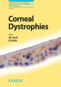
Walter Lisch – Corneal Dystrophies
4.735 ₽
Автор: Walter Lisch
Название книги: Corneal Dystrophies
Формат: PDF
Страницы: 169
Жанр: Медицина
Качество: Изначально компьютерное, E-book
The cornea, basically composed of the epithelium, stroma and endothelium, is the
major refractive organ of the optic system in addition to serving as a mechanical barrier.
The corneal epithelium is the most regular arrangement of stratified epithelium
in the whole human body. The cells, composed of 6– 7 different layers, are tightly
and orderly arranged without intercellular spaces. We know that some corneal dystrophies
are only characterized by the occurrence of epithelial opacities. The contact
lens- induced regression of opacities in epithelial corneal dystrophies can be interpreted
as a contact lens- induced reduction of epithelial layers. As in other connective
tissues, the major portion of the corneal stroma is composed of extracellular matrix
macromolecules which are responsible for the strength and transparency of this tissue.
Some corneal dystrophies are thought to result in part from abnormalities in
corneal stromal cell function. Corneal stromal cells synthesize and degrade matrix
materials during corneal morphogenesis and proper metabolism of such materials is
essential. Stromal corneal dystrophies recur after decades on the graft due to the longterm
transformation of transplant keratocytes into pathological host keratocytes. The
corneal endothelium is a monolayer of hexagonal cells that forms the posterior corneal
surface. An intact monolayer of endothelial cells is essential for the functional
endothelial barrier to preserve a relative dehydration of the stroma and a prerequisite
to corneal transparency. If the integrity of the monolayer is breached, corneal
edema rapidly develops as we can see in some endothelial corneal dystrophies. The
replacement of the posterior cornea, called Descemet’s stripping endothelial keratoplasty,
represents a modern and sophisticated surgical procedure in the treatment of
endothelial corneal dystrophies.
With the revolution in molecular genetics, our understanding of corneal dystrophies
has changed in the last 15 years as disorders have been mapped and the genes
responsible have been identified. Today we know that phenotypic heterogeneity – the
same gene causing different forms of corneal dystrophies – and genotypic heterogeneity
– different genes causing a phenotypically identical corneal dystrophy – do
exist. Research continues to uncover important knowledge on corneal dystrophies.
However, the identification of the gene and mutations in corneal dystrophies can only
be interpreted as a start in the mosaic puzzle for uncovering the complex relationships in the pathophysiological molecular mechanisms. In general, further molecular physiological
examinations and the evaluation of animal models are necessary to precisely
define the essential protein defect in the different types of corneal dystrophy. The
development of a causal therapy for corneal dystrophies must be the big scientific
challenge in the future
Описание
Книга Walter Lisch – Corneal Dystrophies представляет собой современное руководство по наследственным заболеваниям роговицы. Автор подробно разбирает клинические проявления, генетику, диагностику и современные подходы к лечению различных форм роговичных дистрофий.
Издание охватывает актуальную классификацию IC3D, молекулярно-генетические механизмы развития патологии, методы визуализации (конфокальную микроскопию, оптическую когерентную томографию) и дифференциальную диагностику с другими заболеваниями роговицы. Особое внимание уделено хирургическим методам лечения, включая различные варианты кератопластики.
- офтальмологам, специализирующимся на болезнях переднего отрезка глаза
- врачам-генетикам, интересующимся офтальмогенетикой
- ординаторам и слушателям циклов повышения квалификации по офтальмологии
- специалистам, занимающимся трансплантацией роговицы
Только зарегистрированные клиенты, купившие данный товар, могут публиковать отзывы.
Похожие


Другие книги раздела «Офтальмология»
- Samar Basak – Atlas of Clinical Ophthalmology (Second edition)
- J. Bradley Randleman – Refractive Surgery
- Lucio Buratto – Cataract Surgery and Intraocular Lenses
- Jason Barton – Field of Vision. A Manual and Atlas of Perimetry
- Arnab Biswas – Eyelid Tumors
- James A. Katowitz – Pediatric Oculoplastic Surgery (Second edition, 2018)
- Darin R. Goldman – Atlas of Retinal OCT (2018)
- Harry W. Flynn – Endophthalmitis in Clinical Practice (2018)
- Jean-Luc Febbraro – Surgical Correction of Astigmatism (2018)
- Geoffrey J. Gladstone – Oculoplastic Surgery Atlas (2 Volume set, 2018)
- Pablo Artal – Handbook of Visual Optics (2 Volume set, 2017)
- J. Sebag – Vitreous in Health and Disease
- Zia Chaudhuri – Postgraduate Ophthalmology
- Thomas H. Williamson – Vitreoretinal Disorders in Primary Care (2018)
- Nora V. Laver – The Infected Eye
- Mark A. Codner – Eyelid and Periorbital Surgery (Second edition, 2016)
Все книги раздела «Офтальмология» →
Смежные разделы: Медицина на испанском языке · Медицина на немецком языке · Медицина на французском языке · Анестезиология и Реаниматология · Акушерство и Гинекология · Ветеринария · Гастроэнтерология и Гепатология · Дерматология и косметология · Диетология и нутрициология · Иммунология и Аллергология · Кардиология · Лапароскопия и Эндоскопия
Рекомендуем также
- John Westfall – Celestial Shadows. Eclipses, Transits, and Occultations (2015)
- Jeffery Burley – Encyclopedia of Forest Sciences
- Rob Kitchin – International Encyclopedia of Human Geography
- А. Корольченко – Пожаровзрывоопасность веществ и материалов и средства их тушения (Второе издание, 2 Тома)
- F. Bruinsma – Railway Development. Impacts on Urban Dynamics (2008)
- H. Stadtler – Supply Chain Management and Advanced Planning (5th edition)
- A. Janahmadov – Synergetics and Fractals in Tribology (2016)
- Anna M. Fan – Toxicology and Risk Assessment (2015)

Отзывы
Отзывов пока нет.